Shoulder Ligament Anatomy Diagram - 1 - This page is about shoulder anatomy ligaments and muscles,contains soft tissues of the shoulder,shoulder joint;
byAdmin•
0
Shoulder Ligament Anatomy Diagram - 1 - This page is about shoulder anatomy ligaments and muscles,contains soft tissues of the shoulder,shoulder joint;. The shoulder is one of the largest and most complex joints in the body. It is also known as articular ligament, articular larua, fibrous ligament, or true ligament. Shoulder dislocation treatment recovery symptoms. Ligaments of the joints anatomical chart physical therapy. These ligaments are the primary restraint for upward and backward movement of the.
Image result for glenohumeral ligaments right shoulder. Ac joint is a diathrodial joint with a fibrocartilaginous disk. A joint capsule is a watertight sac that surrounds a joint. Because of its location superior to the glenohumeral joint, it acts as a protection to the joint. Use the mouse scroll wheel to move the images up and down alternatively use the tiny arrows (>>) on both side of the image to move the images.
Shoulder Anatomy New York Ny Handsport Surgery Institute from handsurgeonsnyc.com Notice superior labrum and attachment of the superior glenohumeral ligament. Normal anatomy, variants and checklist. (3) a syndesmosis is a joint in which a ligament connects two bones, allowing for a little movement (amphiarthroses). Image result for glenohumeral ligaments right shoulder. Start studying shoulder ligaments and tendons. Although three ligaments protect and surround the shoulder joint. Robin smithuis and henk jan van der woude. Other ligaments in the body include the:
Start studying shoulder ligaments and tendons.
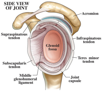
The transverse humeral ligament is not shown on this diagram/caption. Head and neck anatomical chart. 259) may become ossified, transforming the scapular notch into an anomalous bony canal, the scapular foramen. Joints can be grouped by their structure into fibrous, cartilaginous, and synovial joints. We are pleased to provide you with the picture named shoulder anatomy poster size resizing diagram. Normal anatomy, variants and checklist. An image depicting shoulder anatomy can be seen below. Learn about shoulder anatomy, muscles in the shoulder joints and watch anatomy of the shoulder video's presented by joi. Start studying shoulder ligaments and tendons. You can see it enclosing the glenohumeral joint and you can see its attachment on the anatomical you've got the transverse humeral ligament and the coracohumeral ligament. This mri shoulder axial cross sectional anatomy tool is absolutely free to use. There are five major shoulder ligaments that keep the shoulder in place and prevent it from dislocating. Scapular foramen the superior transverse ligament of the scapula (see p.
The primary function of the shoulder girdle is to give strength and range of motion to the arm. Because of its location superior to the glenohumeral joint, it acts as a protection to the joint. The disk has a great variation in size and shape. Normal anatomy, variants and checklist. Although three ligaments protect and surround the shoulder joint.
Shoulder Anatomy from ix-cdn.b2e5.com This page is about shoulder anatomy ligaments and muscles,contains soft tissues of the shoulder,shoulder joint; The primary function of the shoulder girdle is to give strength and range of motion to the arm. Joints can be grouped by their structure into fibrous, cartilaginous, and synovial joints. Anatomy of right shoulder medical illustration medivisuals. Head and neck anatomical chart. We are pleased to provide you with the picture named shoulder anatomy poster size resizing diagram. The distal joint between the tibia and fibula is an example of a. Bones of the upper limb.
Head and neck anatomical chart.
7 draw labelled diagram showing the relations of shoulder joint. Bones of the upper limb. A ligament is the fibrous connective tissue that connects bones to other bones. An image depicting shoulder anatomy can be seen below. One or more ligaments provide stability to a joint during rest and movement. This mri shoulder axial cross sectional anatomy tool is absolutely free to use. Ligaments are soft tissue structures that connect bones to bones. Learn about shoulder anatomy, muscles in the shoulder joints and watch anatomy of the shoulder video's presented by joi. The shoulder is not a single joint, but a complex arrangement of bones, ligaments, muscles, and tendons that is better called the shoulder girdle. The glenohumeral ligaments can be seen here, but they're not really. The transverse humeral ligament is not shown on this diagram/caption. Although three ligaments protect and surround the shoulder joint, most of its stability comes from the powerful muscles and tendons of the rotator cuff. This diagram here just shows the joint capsule itself.
Other ligaments in the body include the: A fold of peritoneum or other membranes. Start studying shoulder ligaments and tendons. Additional stability is provided by: Normal anatomy, variants and checklist.
Shoulder And Axilla Amboss from media-us.amboss.com (3) a syndesmosis is a joint in which a ligament connects two bones, allowing for a little movement (amphiarthroses). Ligaments of the joints anatomical chart physical therapy. Image result for glenohumeral ligaments right shoulder. The conoid and trapezoid ligaments make up the coracoclavicular ligaments. This page is about shoulder anatomy ligaments and muscles,contains soft tissues of the shoulder,shoulder joint; Ligaments are fibrous bands or sheets of connective tissue linking two or more bones, cartilages, or structures together. Although three ligaments protect and surround the shoulder joint. Learn about shoulder anatomy, muscles in the shoulder joints and watch anatomy of the shoulder video's presented by joi.
Although three ligaments protect and surround the shoulder joint.
The shoulder is not a single joint, but a complex arrangement of bones, ligaments, muscles, and tendons that is better called the shoulder girdle. Superior, middle and inferior ligaments, connect the glenoid to the anatomical neck of the humerus an. The multiple ligaments and tendons around the shoulder must be strong to bind the shoulder joints together and encapsulate them in a tough but flexible structure. Home > blog > anatomy > shoulder anatomy: (3) a syndesmosis is a joint in which a ligament connects two bones, allowing for a little movement (amphiarthroses). Use the mouse scroll wheel to move the images up and down alternatively use the tiny arrows (>>) on both side of the image to move the images. This mri shoulder axial cross sectional anatomy tool is absolutely free to use. (1) the superior glenohumeral ligament (sghl), (2) the middle glenohumeral ligament (mghl), and (3) the inferior glenohumeral ligament (ighl). A joint capsule is a watertight sac that surrounds a joint. Although three ligaments protect and surround the shoulder joint. These ligaments are the primary restraint for upward and backward movement of the. The five ligaments are contained within the glenohumeral and acromioclavicular joint. Start studying shoulder ligaments and tendons.
Although three ligaments protect and surround the shoulder joint shoulder anatomy diagram. The primary function of the shoulder girdle is to give strength and range of motion to the arm.